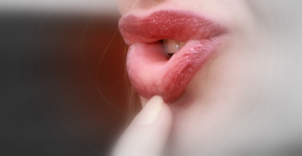
What is the inside of your lip called?
The mouth edge of most vertebrates is formed by the soft, malleable anatomical structure known as the lip, which is made up of a muscle layer, connective tissue, and surface epidermis (skin).
Sebaceous (oil) glands, hair, and sweat glands can all be found in the man's outer skin. The vermilion border—a term used to describe the crimson skin that covers the lips' edges—is densely dotted with delicate nerve endings. The reddish skin serves as a barrier between the inside mucous membrane and the exterior, hair-bearing tissue. A mucosal membrane that is wet along the inside surface of the lips. The inner surface is significantly thicker and has sebaceous glands and tiny projections known as papillae on newborn newborns. These structural modifications appear to facilitate sucking. The orbicular oris muscle, which surrounds the aperture, supplies the majority of each lip's material. The numerous changes in shape and expression of the lips are made possible by this muscle and others that extend out into the cheeks.
Herpes simplex, which causes fever blisters or cold sores, and leukoplakia, which causes white spots that may be precancerous, are two diseases that specifically affect the lips. On the vermilion edge of the lower lip, ulcers in older men are commonly malignant. Additionally, the weather's excessive drying, chemical irritants, an infection's inadequate moistening, or an antibiotic's response might cause the borders to crack and swell.

Oral mucosa
The mucous membrane that lines the interior of the mouth is called the oral mucosa. The "oral epithelium" is a stratified squamous epithelium that is made up of lamina propria, a connective tissue beneath it. The mouth cavity has occasionally been compared to a mirror that shows how healthy a person is. Alterations in the oral mucosa, which lines the mouth, are considered changes symptomatic of disease and can indicate local consequences of long-term alcohol or cigarette use as well as systemic disorders like diabetes or vitamin deficiencies. Comparatively speaking to the skin, the oral mucosa tends to recover more quickly and with fewer scars. Though the fundamental process is uncertain, research points to extracellular vesicles as potential players.
Classification
Based on function and histology, there are three basic kinds of oral mucosa:
- Nearly every other area of the oral cavity also has nonkeratinized stratified squamous epithelium as mucosal lining, including the:
- Between the buccal and labial mucosae is a layer called the alveolar mucosa. It is a brighter red, smooth, glossy structure with numerous blood arteries, and no rete pegs are holding it to the surrounding tissue.
- Buccal mucosa, or lining mucosa, is the inside lining of the cheeks.
- Lips' internal lining, or labial mucosa, is a kind of mucosa.
- On the dorsum of the tongue, hard palate, and connected gingiva is a keratinized stratified squamous epithelium known as the masticatory mucosa.
- Nerve terminals for general sensory reception and taste perception are found in the areas of the taste buds on the lingual papillae on the dorsal surface of the tongue.
Structure
The surface stratified squamous epithelium and the deeper lamina propria make up the two layers that make up the oral mucosa. The epithelium in keratinized oral mucosa has four layers:
- Stratum basale (basal layer)
- Stratum spinosum (prickle layer)
- Stratum granulosum (granular layer)
- Stratum corneum (keratinized layer)
Depending on the area of the mouth, the oral mucosa may be keratinized or nonkeratinized. To create the stratum corneum, keratinocytes must differentiate into nonvital surface cells, a process known as keratinization. In reaction to trauma, the nonkeratinized epithelium can change into a keratinizing type, which results in hyperkeratinization. Chronic physical trauma might cause additional hyperkeratinization in even keratinized tissue. The lamina propria is a thick and papillary layer of fibrous connective tissue. In the thick layer of the lamina propria, the submucosa may or may not be present. An ordinary variation of the oral mucosa is Fordyce spots. At the junction of the lamina propria and the oral epithelium is the basal lamina.
Functions
Eating, drinking, and talking continually generate mechanical stress in the mouth environment. The mouth must be able to respond fast since it is likewise susceptible to abrupt changes in pH and temperature. The body only has one location where taste may be experienced: the mouth. The oral mucosa must perform a variety of specialized duties as a result of these special physiological characteristics.
Protection
Physical protection of the underlying tissues from mechanical forces, germs, and toxins in the mouth is one of the major purposes of the oral mucosa. The hard palate and gingivae are firmly attached to the keratinized masticatory mucosa. It makes up 25% of the whole oral mucosa. By fending against the loading forces generated during mastication, it supports the tissues below. When speaking and chewing, the lining mucosa in the cheeks, lips, and mouth floor can move around to make room. It facilitates unrestricted movement of food during mastication while also protecting the underlying tissues from damage. It makes up 60% of the oral mucosa.
Secretion
The oral mucosa's main secretion is saliva. It serves a variety of purposes, such as lubrication, pH buffering, and immunology. Resting is the keyway to maintaining saliva's lubricating and antibacterial properties, which flush the mouth and clear harmful substances and oral waste. Numerous antimicrobial proteins found in saliva assist to shield the oral ecology against pathogenic agents. The presence of substances in the saliva that operate as a defensive mechanism includes lysozyme, lactoferrin, salivary peroxidase, myeloperoxidase, and thiocyanate concentrations. Three primary pairs of salivary glands (parotid, submandibular, and sublingual) as well as numerous smaller salivary glands release saliva. As it includes the enzyme amylase, which converts carbs into sugars, it also helps the food's early chemical digestion.
Sensation
The extensive innervation of the oral mucosa makes it exceptionally sensitive to pain, touch, temperature, and taste. Sensations in the mouth are processed by some cranial nerves, including the trigeminal (V), facial (VII), glossopharyngeal (IX), and vagus (X) nerves. There is specialized mucosa on the tongue's dorsum. Around 15% of oral mucosa is made up of this, which also contains taste buds that enable taste. Mouth reflexes like swallowing, gagging, and thirst are also started here.
Thermal regulation
Some animals, such as dogs, rely on panting to control their body temperature since their only location for sweat glands is on their feet, a factor that is relevant in humans but not in other animals.
Conclusion
The oral mucosa, the mucous membrane lining the interior of the mouth, is the term used to describe the interior of the lip. The vermilion border acts as a partition between the inside mucous membrane and the outside, hair-bearing tissue. The lips are composed of a muscular layer, connective tissue, and surface epidermis. The oral mucosa carries out many specific tasks, including guarding the underlying tissues against physical stress, pathogens, and toxins in the mouth, as well as enabling taste perception and reacting swiftly to pH and temperature changes. Alterations in the oral mucosa can also be a sign of systemic diseases like diabetes or vitamin deficiencies as well as local effects of long-term alcohol or cigarette use.